Comparing Prostate Cancer Diagnostic Methods: MRI Scan or Biopsy?
Prostate cancer is a disease that affects the prostate, a small gland located between the penis and the bladder in males. This article explores the roles of MRI scans and prostate biopsies in the diagnosis and management of prostate cancer.
MRI Scans
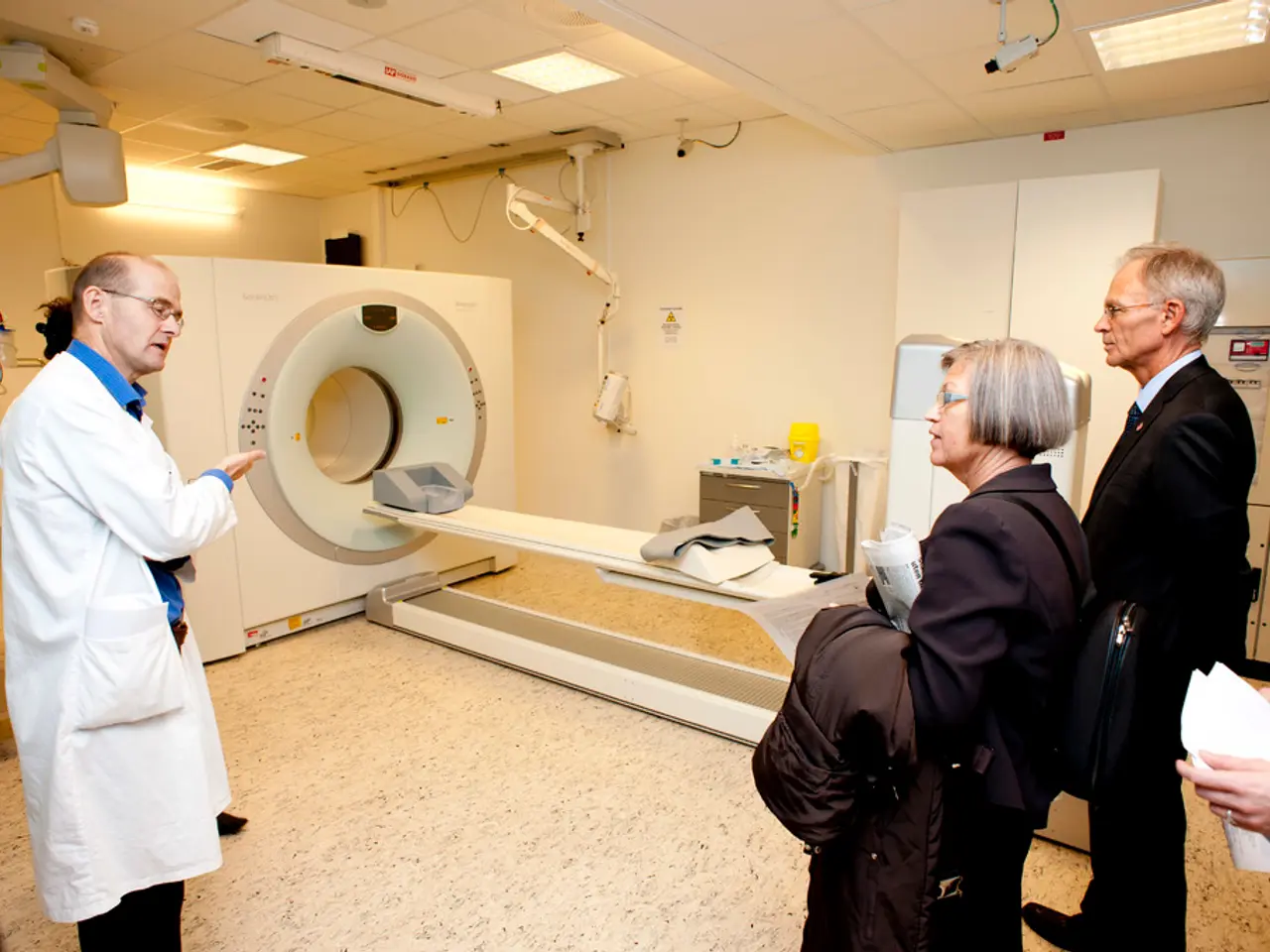
MRI scans are non-invasive imaging methods primarily used before biopsy. They help identify suspicious areas within the prostate that may harbor clinically significant cancer. MRI scans use strong magnetic fields and radio waves to create images of the inside of the body, providing a detailed view of the prostate.
MRI scans improve detection accuracy by targeting these suspicious lesions, thereby reducing overdiagnosis of low-risk cancers and minimizing unnecessary biopsies. MRI also allows visualization of prostate anatomy and tumor characteristics, which assists in planning personalized treatment and active surveillance strategies. Additionally, MRI can guide radiation therapy in real time, improving treatment precision and reducing side effects.
Prostate Biopsies
Prostate biopsies involve extracting tissue samples from the prostate for histological examination to confirm and grade cancer. While biopsy remains the diagnostic gold standard, it is an invasive procedure with risks such as bleeding and infection.
Traditional biopsy without MRI guidance may miss significant tumors or detect insignificant ones, sometimes leading to overtreatment. However, MRI-informed biopsies, which use MRI findings to target suspicious areas, have enhanced the diagnostic yield for clinically significant cancers while limiting detection of indolent tumors.
Benefits and Limitations
Benefits of MRI:
- Non-invasive imaging.
- Improves detection of significant cancers.
- Reduces overdiagnosis of low-risk cancers.
- Guides precise biopsy targeting.
- Enables real-time adaptations during radiation therapy to protect healthy tissue and minimize side effects.
Benefits of Prostate Biopsy:
- Provides definitive cancer diagnosis through tissue analysis.
- Determines cancer grade and pathology essential for risk stratification and treatment planning.
- MRI-targeted biopsies have improved accuracy over systematic biopsy alone.
Limitations of MRI:
- Cannot confirm cancer alone, only suggests suspicious areas.
- May miss some small or less conspicuous tumors.
- Access and cost can be limiting factors.
Limitations of Biopsy:
- Invasive procedure with risks.
- Systematic biopsy can miss tumors or overdiagnose low-risk disease.
- Sampling errors possible without MRI guidance.
How They Are Used Together
The current standard typically involves performing an MRI first to identify suspicious lesions. MRI findings guide targeted biopsies to the most concerning areas. This combined approach improves detection of significant prostate cancer while avoiding unnecessary biopsies and overdiagnosis.
After diagnosis, MRI is used in active surveillance to monitor tumor stability, potentially reducing the need for repeated biopsies unless MRI or clinical changes suggest progression. MRI also plays an increasing role in guiding and improving the precision of radiation therapy.
In summary, MRI enhances the diagnostic accuracy and management of prostate cancer by refining biopsy targeting and treatment planning, while biopsy remains essential for definitive diagnosis and pathological grading. Their combined use optimizes patient outcomes by balancing accurate detection with minimization of invasive procedures and overtreatment.
It's important to note that symptoms of prostate cancer may include frequently needing to urinate, weak urination streams, and blood in the urine. Some people may experience soreness, bleeding, and discolored semen for several weeks after the procedure. Prostate biopsies are often invasive, involving the insertion of a hollow needle into the prostate.
[1] Khan, S. A., & Ahmed, H. (2020). Prostate MRI: Techniques and Applications. Springer.
[2] Kattan, M. W., & Ahmed, H. (2017). Prostate MRI: From Diagnosis to Treatment Planning. Springer.
[3] Böhm, M., & Ahmed, H. (2016). Prostate MRI: From Diagnosis to Treatment Planning. Springer.
[4] Ahmed, H., & Böhm, M. (2015). Prostate MRI: From Diagnosis to Treatment Planning. Springer.
[5] Ahmed, H., & Böhm, M. (2014). Prostate MRI: From Diagnosis to Treatment Planning. Springer.
Read also:
- Overweight women undergoing IVF have a 47% higher chance of conceiving naturally post-weight loss
- Bonsai Trees from Evergreen Species: Exploring Growth Characteristics & Distinct Qualities
- What temperatures may make walking your canine companion uncomfortable?
- Alcohol consumption and the connection to esophageal cancer: An exploration of links and potential hazards


